Vision question

Greg Bockelman

Touchdown! Greaser!
Joined
Feb 23, 2005
Messages
11,093
Location
Lone Jack, MO
Display Name

Display name:
Greg Bockelman
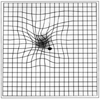
Help me figure out what vision problem is being tested for here.

When you are looking at a spot in the middle of a card with a grid on it, the lines appear wavy or there are gaps in the lines. What condition is this, and what might be a treatment for it?
 
Greg, that can be a lotta stuff, or it can be nothing. The key is to get a good dilated exam (the tool the opthalmologist uses, an Indirect Opthalmoscope, can see wayyy out to the retinal edges with a dilated pupil. Because, if anything needs done, that' how he's going to find it.

It can be nothing. It can be spotty dengeneration of various types. It can just be plain ol' astigmatism ("rumply" retina).

I really hate the waste of the rest of the day when the opthal. peers into my brain (the retina is a piece of the brain) but it's worth it periodically.

"don't panic, lads!"
 
Is this a problem you are experiencing, I mean right now ?

Like this:
Amslergrid_patientAMD.gif


Or more like this:

6613_FIG.%2017.4.jpg


Either way, get this checked out soon, as in tomorrow morning soon. Dilated Fundus Exam (DFE). Can be a couple of things, some good, some not so good.

Any flashing lights ?
 
I always do the picture rather than the dilated exam. It costs more since it's not covered by insurance but I don't like the aftereffects of the eye drops. Besides, then the doctor can sit there and go over the pictures with me and compare them to last year.
 

My wife has that on a magnet on the refridgerator. Her retina specialist gave it to her. She's had problems in the past with PVD and floaters, large ones. But no retinal detachment.

IIRC if u look at it with one eye, at the right distance, you may find a hole where the grid is not visible. That should be were the optic nerve connects. With two eyes, the other eye fills in the hole. The brain is so smart about stitching the two visual clues from each eye together to get one complete picture.
 
Mari, the Amsler graph paper is complimentary (and additional) to the dialated exam, not a replacement. You can't accurately report on the edges of your Visual Fields, which is where detachments start..... sigh.
 
Mari, the Amsler graph paper is complimentary (and additional) to the dialated exam, not a replacement. You can't accurately report on the edges of your Visual Fields, which is where detachments start..... sigh.
I wasn't talking about the paper graph. I was talking about the picture they can take of the inside of your eye showing the retina, the optic nerve, the blood vessels, etc. My eye doctor has had this camera (or whatever it is) for a few years now. She talked me into getting the picture as part of my regular exam a couple years ago since I kept refusing the dilation and now I do it every year. I don't know if this is the exact unit she uses but shows what I mean by "pictures".

http://www.totaleyecare.com/Optomap.htm
 
Mari, that is exactly it. I was trying to find out what condition it was supposed to diagnose.

It is a screening tool for a number of retina problems.

Most commonly, this is given home with patients who have diabetes and are at risk for complications related to that. Also, older patients with age related retinal degeneration are given this chart as a self-test to pick up on things happening to the retina. Also, patients who are quite near-sighted (myopic) are at an increased risk of experiencing retinal detachments, at times they will be given a Amsler grid to self-test for that happening. In all of the stuff that can happen to the retina, acting earlier rather than later is of great benefit.
 
Old Thread: Hello . There have been no replies in this thread for 365 days.
Content in this thread may no longer be relevant.
Perhaps it would be better to start a new thread instead.
Back
Top